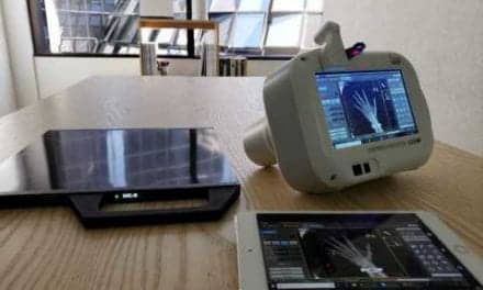
A new study by the University of Texas MD Anderson Cancer Center concluded that tomosynthesis is highly accurate for detecting positive margins intra-operatively, compared to traditional 2D imaging with extensive processing, in breast cancer patients undergoing segmental mastectomy.
Statistics indicate that nationally, 20 to 30% of these patients need to undergo re-excision in order to obtain negative margins. The results of the study indicate that intraoperative tomosynthesis is more accurate with greater specificity than standard extensive processing at identifying positive margins, and is also a simpler process that is less likely to lead to unnecessary additional tissue excision.
The study included 99 patients undergoing segmental mastectomy for breast cancer, and compared the clinicopathologic features of specimen margin status using standard extensive processing and intraoperative specimen tomosynthesis using the Mozart Specimen Tomosynthesis System from Kubtec, based in Stratford, Conn. The institution’s established SEP, while oncologically effective, is a multistage process involving 2D x-ray imaging of the intact specimen, gross evaluation of the specimen, then sliced into 5mm slices, and review of the multiple 2D x-ray images of the sliced specimen reviewed intraoperatively by a radiologist.
Definitive margin status of each specimen was based upon permanent pathology evaluation.


