By Aine Cryts
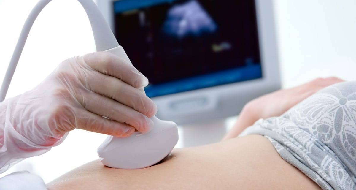
Automated breast ultrasound (ABUS): Stamatia Destounis, MD, attending radiologist at Rochester, N.Y.’s Elizabeth Wende Breast Care, highlights this as one of the most widely used ultrasound technologies in imaging today. “This newer technology improves upon some of the limitations of traditional handheld ultrasound, such as operator dependency and examination time,” she tells AXIS Imaging News.
Destounis is excited about automated breast ultrasound for three reasons:
- The scans provide physicians with a 3-D volumetric image of the entire breast.
- Automated ultrasound has been shown to require a shorter examination time.
- Since the transducer used in automated ultrasound systems scans the breast automatically, operator dependency can be minimized.
ABUS, which creates 3-D images of the breast, may be especially useful as a complement to traditional mammography to identify developing tumors in women with denser breast tissue.
Artificial intelligence (AI), is another technology that Destounis describes as a“hot topic” in breast imaging and breast ultrasound, mostly in the use of computer-aided detection (CAD). “Multiple studies have shown promise with this emerging technology, demonstrating improved reader accuracy in the detection of breast lesions on ultrasound, as well as for ultrasound CAD to aid in image-review times, [thus,] allowing radiologists to be more time efficient,” says Destounis, who is also clinical professor at University of Rochester Imaging Services.
Then there is shear-wave elastography (SWE), a relatively new technology that uses acoustic radiation to evaluate the elasticity of soft tissue, is also proving useful at distinguishing benign from malignant breast lesions.1,2 This may help to reduce unnecessary biopsies of benign lesions,” says Destounis. Ongoing study of AI with elastography—for example, using radiomic features—may improve diagnostic performance even more, she says.
Aine Cryts is a contributing writer for AXIS Imaging News.
References
- Sebag F, Vaillant-Lombard J. Berbis J, Griset V Henry JF, Petit P, Oliver C. Shear wave elastography: a new ultrasound imaging mode for the differential diagnosis of benign and malignant thyroid nodules. J Clin Endocrinol Metab. 2010;95(12):5281-8. doi: 10.1210/jc.2010-0766.
- Youk JH, Gweon HM, Son EJ. Shear-wave elastography in breast ultrasonography: the state of the art. Ultrasonography. 2017;36(4):300-309. doi: 10.14366/usg.17024.


