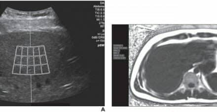
There have been many technologic advances in diagnostic sonography, which affect its clinical use and economic impact. This overview provides a description of those recent developments that have the potential to make major clinical and economic impact. These include improvements in image processing, system miniaturization, and the potential use of contrast sonography. The development of clinical applications from these technical innovations has implications regarding training and practice, as well as economic factors involved in the practice of diagnostic sonography.
Image Processing
Recent technological advances have improved image resolution as well as allowed diagnostic sonography to portray physiologic processes. These technologic advances include the use of harmonics and pulse inversion for improvement of textural resolution, as well as for depiction of physiologic processes such as organ perfusion and function with the use of sonographic contrast agents. The addition of the use of contrast will afford assessment of organ vascularity and flow. From these parameters, one can infer a variety of features such as normal and abnormal function, benign vs malignant vs inflammatory processes.
The principle of harmonics is that the signal-to-noise ratio can be improved by listening at a multiple of the fundamental frequency. Although the signal returned is quite small, it can be amplified and has better signal-to-noise ratio than the fundamental frequency. Pulse inversion also improves the signal-to-noise ratio with the use of microbubble contrast. In this technique, an inverted duplicate of the emitted pulse is sent. Where there is in-phase construction, the pulses cancel each other whereas when a microbubble is encountered, it causes the bubble to resonate and the out-of-phase signal is amplified. This has major applications for the use of contrast in assessment of enhancement kinetics, which reflects the perfusion within a tissue area.
Another improvement in signal processing is more extensive use of 3-D imaging. There are several definite clinical applications of 3-D imaging, such as surface rendition of fetal facial features. In addition, 3-D imaging has a major advantage in depicting tissue volumes. This is helpful, for example, in following fibroid enlargement and thyroid nodule enlargement, and in depicting spatial relationships of masses to other structures. 3-D color Doppler sonography depicts overall vascularity and thus can show branching patterns of vessels within masses.
A major improvement in signal processing has occurred with electronic compounding. This application electronically pulses the subelements of the transducer and images structures at multiple angles. Where there is no image construction, scatter is effectively reduced, leaving an improved image. This technique will have major applications in improved soft tissue evaluation of structures such as the breast where the radiographically dense breast is still a difficult structure for evaluation and detection of breast carcinoma. Predictably, compounding will also have many applications for musculoskeletal disorders.
System Miniaturization
Technological advancement will improve miniaturization of equipment. Scanners now the size of or smaller than laptops are available. These will become extensively utilized in emergency departments and trauma units for the depiction of major (nonsubtle) abnormalities, such as the presence of free intraperitoneal fluid. They will also be helpful in the intensive care unit for bedside placement of central venous lines and depiction of pleural fluid. Thus, it is envisioned that these small portable units will assume a major role in guided procedures, such as percutaneous placement of catheters that can be performed at bedside.
The more extensive utilization of smaller mobile sonography units has implications for the quality of sonograms performed. Sonography, by its nature, requires dedication to full and complete understanding of anatomy and normal and abnormal physiologic processes. Erroneous diagnosis and poor use of diagnostic sonography can result in untoward effects of mismanagement and mistreatment. Thus, it is important that certain governing societies such as the American Institute of Ultrasound in Medicine adopt accreditation processes and that users dedicate themselves to continuous medical education in diagnostic sonography.
This overview should provide the reader with the latest applications of diagnostic sonography. As diagnostic sonography continues to grow and permeate throughout many medical disciplines, it is imperative for quality patient care that the user be educated and able to appropriately use this highly versatile diagnostic modality.
Arthur C. Fleischer, MD, is professor of radiology and radiological sciences and chief of diagnostic sonography at Vanderbilt University Medical Center, Nashville, Tenn.
Related Links:


