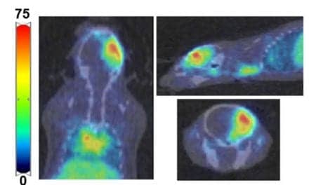
The field of nuclear medicine is poised for an exciting leap forward into the next century. Positron emission tomography (PET) is the white knight-establishing new standards of cancer detection and staging through an accumulation of clinical data and technical advances as well as political gains that are gradually changing third-party and Medicare reimbursement for the procedure from a trickle into a steady stream.
PET has been a technique in search of indications since it became available in the 1970s. The cost of acquiring a scanner and a cyclotron to generate the necessary radiopharmaceuticals often exceeded $4 million-not including personnel and annual maintenance.
Despite its ability to image metabolism rather than anatomy, PET was prohibitively expensive for most sites. Those institutions that did acquire PET, however, doggedly persevered in efforts to establish a clinical database showing that PET could prove invaluable in examining the brain, heart, and whole body in new ways. Although change and clinical acceptance have been slow in coming, PET has now arrived.
“Many developments have occurred in the last 18 months that have had a major impact on PET,” according to R. Edward Coleman, MD, professor and vice-chairman of the Department of Radiology and director of the Division of Nuclear Medicine at Duke University, Durham, NC. “In January 1998, the Health Care Financing Administration (HCFA), which sets reimbursement policy for Medicare, approved coverage for two new PET indications-evaluation of solitary pulmonary nodules and the staging of lung cancer. Following a town hall meeting just 1 year later, HCFA approved an additional three oncology-related indications-detection and localization of recurrent colorectal cancer, staging and characterization of Hodgkin and non-Hodgkin lymphoma, and identification of metastases in recurrent melanoma.”
PRIVATE SECTOR PAVES WAY
Coleman finds the recent HCFA-approved PET indications in oncology somewhat ironic in the sense that the federal government is following the lead of the private sector instead of the other way around. The Blue Cross/Blue Shield Technology Evaluation Center reviewed PET in the spring of 1997 and concluded that it should be covered when used in diagnosing and staging lung cancer-a year before HCFA reached the same conclusion. Other carriers have begun to follow suit and reimbursement is finally flowing on a case-by-case basis if not as part of a broad, global coverage policy.
“Perhaps the single most important factor underlying coverage by Medicare and private third-party payors is the critical mass of clinical data that has developed in the past 5 years,” explains Ruth Tesar, president of the Institute for Clinical PET, Foothill Ranch, Calif, and vice president/general manager of a company that provides a network of radiopharmaceutical services. “All the research targeting the clinical indications that really make a difference in patients’ lives has finally come to the fore. Referring physicians are beginning to acknowledge that PET accuracy for the recently approved oncologic applications is in the 90% range-superior to CT with contrast as well as MRI-and that has caught their attention,” she states.
Equally important to achieving reimbursement, Tesar notes, has been the personal interest and active participation of Senator Ted Stevens (R-Alaska) who has repeatedly urged the Secretary of Health and Human Services and the director of HCFA to make coverage available.
“Without his personal intervention with Secretary Shalala, I do not think we would be looking at the approval of these new indications,” she acknowledges.
FROM HEART TO WHOLE BODY
The advance of PET from cardiac perfusion studies into oncologic imaging began only in 1992-93 when whole-body scanners with adequate resolution and improved software for image acquisition, processing, and display became available, according to Peter E. Valk, MD, medical director of PET at Northern California PET Imaging Center, Sacramento, Calif. “I think there is a misconception that we have been using PET in oncology for a long time,” Valk says. “It is like ultrasound, however, which was around for a long time before the machines were built that actually made it a clinical reality. The same thing applies to PET oncology.”
Better data have arrived with next-generation PET systems. Valk, Coleman, and others point to prospective studies that have demonstrated the clinical efficacy of PET imaging in the diagnosis and staging of recurrent colorectal cancer, recurrent ovarian cancer, cancer of the pancreas, abdominal and pelvic lymphoma, and metastases from lung cancer, esophageal cancer, and malignant melanoma. PET is more sensitive and specific than anatomic imaging modalities, thereby permitting more appropriate treatment selection.
From a managed care perspective, the growing body of literature clearly demonstrates that the preoperative use of fluorine 18-labeled deoxyglucose (FDG) PET scanning helps patients avoid unnecessary surgeries and biopsies. In the preoperative staging of recurrent colorectal cancer and metastatic melanoma, for example, FDG PET has been shown effective in avoiding surgery for nonresectable tumor because it can display previously unsuspected sites of active cancer. The bottom line, in a sense, is that while FDG PET scans cost more than CT or MRI, they can save patients and hospitals substantial money in the longer term.
“We are talking about patients who would not benefit from having a tumor removed because it has already spread to other parts of the body that are not operable,” Coleman says.
Perhaps more than cost-effectiveness, quality of life is a factor. “Unnecessary surgery costs the patient a lot more than money, in that if you have got only a year to live, you do not want to spend it having a useless operation and then trying to recover from it,” Valk adds.
PET ON THE RISE
The number of centers actively doing clinical PET has grown as well, Tesar reports. As recently as 1995, there were 80 PET sites in the United States, of which 20 to 30 did research only. Growth was limited because reimbursement was limited when it was available at all and because it cost too much to install the technology and operate a PET program. The number of clinical PET sites has grown since then because of the convergence of three developments: the use of collimators on gamma cameras, the addition of coincidence detection capability to dual-headed gamma cameras, and the nationwide availability of FDG through commercial networks of pharmaceutical services companies, eliminating the need to buy a $2 million cyclotron.
Today there are 200 sites performing clinical PET imaging with dual-use gamma camera technology, Tesar reports.
“Sites that use the coincidence cameras are not doing the same volumes as those with dedicated PET scanners because most use them for conventional nuclear medicine studies at least half the time,” she explains. “They are learning how best to employ the modality and I would imagine that with HCFA’s latest approval of new indications, they are apt to get very busy in the near future.”
Gamma camera manufacturers have also made attenuation correction available for PET imaging to increase their accuracy in oncologic applications, according to Coleman.
“There have been a lot of changes in the technology that are resulting in improved imaging, which in turn results in more accurate diagnoses,” he notes.
Valk attributes decreased equipment costs with helping to further the diffusion of PET technology. “I believe that the gamma cameras designed to perform positron imaging do a pretty good job in specific circumstances, such as the lung and head and neck,” he says. “They are not as effective as the dedicated PET scanners in the thicker, more solid parts of the body, but they are substantially less expensive. What is also happening is that dedicated devices are becoming less expensive, and I believe that within 2 years, we will see dedicated PET scanners that cost no more than a high-end gamma camera.”
Indeed, FDA-approved PET systems can now be bought for about $800,000-a far cry from the roughly $2.5 million spent on Valk’s machine in 1992. “Today, a top-of-the-line dedicated PET scanner costs around $1.5 million-about what a hospital might spend for an MR imager,” he notes. “I believe that within 2-3 years, you will be able to buy a high-end PET scanner for under $1 million.”
Valk and other experts expect a reduction in PET scan costs as machines become less expensive to buy and operate. They also predict that the number of scans performed each day will increase because of the growing number of approved oncologic indications. Together with the expanded availability and affordabilty of FDG, the anticipated increase in the number of procedures performed should contribute to further cost efficiency at PET sites.
As nuclear medicine departments around the country begin to realize that expanded coverage for PET scanning could turn an expensive research-only technology into a clinically useful and profitable oncology procedure, “To buy or not to buy?” will become the question.
ADDING PET
Tesar believes that physicians and hospital administrators should formulate clear ideas on what their departmental product line ought to include. Upgrading an existing gamma camera with coincidence detection capability might work in some instances, but prove less satisfactory in others. She and many experts agree that PET imaging represents a new gold standard in a growing number of oncologic situations.
“A PET scan should be done any time a patient is faced with surgery for colorectal cancer or melanoma,” she states, “because so many times more disease is found.”
Tesar is not alone in suggesting that PET scanning become the standard of care in specific oncologic situations. Because PET imaging is available in Sacramento, the Kaiser Foundation Health Plan there has written it into its protocol for evaluating pulmonary nodules. Similarly, she indicates that Physicians Reliance Network acquired a PET scanner. PRN works with the Texas Oncology Practice Association (TOPA), representing 200-300 oncologists, and is beginning to incorporate PET into practice parameters.
“PET is revolutionizing nuclear medicine,” Tesar says. “It is changing the way the field is perceived by physicians. Nuclear medicine has never been able to be the choice for oncology, but now it will be.”
Beyond the excitement generated by HCFA’s latest indication approvals and the concomitant increase in the reimbursement stream, Coleman and Valk agree that the evaluation of treatment effectiveness could be PET’s next frontier.
They explain that when tumors are treated with radiation and chemotherapy, some disappear entirely while many others shrink to varying degrees and then stop. CT and MRI cannot discern whether the remaining tissue consists solely of necrotic matter or active tumor. Fine-needle biopsy is only a partial answer, because a finding of scar tissue is no guarantee that living cancer cells were not present.
“The metabolic characteristics of PET do not suffer from this drawback,” Valk explains, “because scar tissue is not metabolically active.”
ASSESSING TREATMENT
PET technology may help decide a related issue in oncology: response to chemotherapy. How long and what type are quality of life and economic issues that have not been fully investigated. A technology that could rapidly document tumor response or the lack of one might spare the patient and simultaneously control costs.
“Preliminary studies suggest that we might not have to give patients 4-6 cycles of chemotherapy to determine its efficacy,” Valk adds. “We may be able to administer one or two cycles, then do a PET scan and be able to see whether the cancer has responded sufficiently to justify continuing.”
Valk points to the fact that many people talk about the cost of diagnostic imaging modalities and surgery, but few studies ever get conducted.
“There has been very little discussion of the fact that in the United States, it has become practically illegal to die of cancer without being on chemotherapy-and there has been no discussion of what a cycle of chemotherapy costs,” Valk notes. “No one has even started to look at cost-efficacy in this area. It is a huge operation that rolls along at enormous cost-and no one evaluates it. Ultimately, PET scanning may help us determine how well we treat a patient as well as how cost-effectively.”
Coleman, Valk, and their colleagues believe that HCFA’s next round of approved PET indications will include brain tumors and head and neck cancers.
“I am not quite sure why HCFA decided on the three of the five indications that they did, but we will continue to provide them with data on brain tumors and on head and neck cancers, and I would certainly think that those are very likely to be covered in the near future,” Coleman says.
Perhaps more significantly in the long term, PET experts hope to obtain global coverage for PET rather than continuing to gain piecemeal approval, tumor by tumor.
“The concept is identical for all oncology applications,” Tesar says, “whether it is breast cancer or recurrent colorectal cancer. Perhaps what you do as a result of the scan will vary, and maybe what brings the patient in to have a PET scan may be different, but the way PET works does not change from tumor to tumor. We are seeking metabolic activity, and when it is present, we are 90% certain that we are seeing tumor. Why differentiate by cancer type?”
USE IN ALZHEIMER’S
Lest the medical and managed care communities conclude that PET use in oncology represents the only major news in nuclear medicine, Tesar points to the technology’s use in Alzheimer’s disease.
“We know that PET can help us detect Alzheimer’s disease at a very early stage,” she explains. “Because new drugs are now available, patients may actually be able to delay the onset of crippling symptoms. By selectively using PET for at-risk patients, it might be possible to delay the need for out-of-home care-which could save millions of dollars.”
With reimbursements beginning to flow for PET, experts hope that a ripple effect will benefit the entire field. That may already be happening with monoclonal antibody and peptide products and techniques that are increasingly refined and may soon find applications from cancer to blood clots.
“There are some very exciting data about monoclonal antibody applications in cancer treatment, with new data from phase 3 clinical trials showing efficacy in treating lymphoma,” Coleman notes. “There are also approved new compounds showing that nuclear medicine techniques are very accurate in detecting blood clots. We may soon see nuclear medicine playing a role in the detection of venous thrombosis,” he adds.
“PET continues to gain momentum,” Tesar says, “and with it, nuclear medicine as a whole. We look forward to a bright future.”
?
Sheldon M. Stern is a freelance medical writer in Irvine, Calif, and a contributing writer for Decisions in Axis Imaging News.


