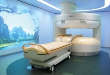
High image quality, low cost technology, and greater patient comfort features are just a few of the demands today’s MRI machines must meet.

With Philips? Ambient Experience, patients relax during an MRI exam while viewing nature scenes of the beach or desert.
Today’s health care organizations seek out particular MRI equipment to meet the needs of patients and the business goals of the organization. Not every patient needs to be sequestered in an expensive, claustrophobia-producing, closed design, whole body MRI machine. For example, a high school athlete with a sprain can keep his head out of the machine while getting his ankle scanned using an MRI for limbs only. And for those patients who do require an MRI beyond the limbs, an open MRI is now possible, allowing for “views” of the beach, Africa, and more.
GE recently introduced the Optima MR430s musculoskeletal “extremity” scanner allowing for targeted scanning of the limbs. Patients sit and only targeted anatomy—an arm or leg—goes inside the scanner. As such, it meets a growing demand for comfort since its innovative design makes technology surprisingly more patient-friendly as compared with traditional MRI systems. It also meets a business demand, as the Optima MR430s is an ideal adjunct for a busy radiology department due to its cost-effectiveness. The new GE extremity scanner also features a smaller footprint versus traditional MRIs, and easier installation, and it still produces high-quality images at a low total cost of ownership. Its use can allow the full-body MRI to remain available for patients who require it, thereby relieving backlogs and increasing department efficiency.
For those patients who do require a full MRI such as of the head, shoulders, abdomen, or chest cavity, the open design of the Philips Panorama High Field Open (HFO) MRI with Ambient Experience is an option. So instead of the whirling sound inside an enclosed tube, the newly revised sensory experience integrates architecture, design, and technologies, such as lighting and sound, to allow patients to personalize their environment and experience a more relaxing atmosphere. Animation selections on the wall are accompanied by soft lighting and music, allowing “escapes” to areas such as the coastline or mountains. For children, a cartoon scene is also available.
Another system that makes the MRI exam easier for pediatric patients is Toshiba’s Vantage Atlas® MRI. This system includes features specifically designed to improve patient comfort. Its Pianissimo technology reduces noise by up to 90% during MR exams, allowing for a quieter, more comfortable exam. It also includes a short, open bore that provides a feeling of openness that reduces claustrophobia. Furthermore, the system includes Atlas integrated coil technology, which reduces the need for repositioning patients during an exam.
Evidence of the value of all of this improved MRI technology comes from reports from the institutions that have benefited from it.
Concentrated Quality

Philips? Ambient Experience.
William B. Morrison, MD, director, Division of Musculoskeletal Radiology at Thomas Jefferson University, Philadelphia, discussed the use of the Optima MR430s musculoskeletal extremity scanner. The university was an early adopter of this technology, which GE Healthcare purchased from the ONI company in 2009. Since then, the ONI Extreme has been reengineered and was launched by GE as the Optima MR430s in February.
“As musculoskeletal radiologists, we want high-quality scans,” said Morrison when asked what prompted his department to purchase the extremity scanner that was the predecessor to the Optima MR430s. Morrison noted a host of issues regarding anatomy, MRI function, and patient compliance, supporting the idea that whole body MR scanners are not necessarily the best choice for imaging the extremities. “Whole body scanners are retrofit for the extremities. They are optimized for imaging the brain, and the thoracic, abdominal, and pelvic areas. They are not made for the extremities. They can be good for the knees, but the ankles and feet have always been an issue because the coils don’t accommodate the anatomy very well.
“Upper extremities have been a problem as well,” he said. “Shoulders are positioned at the edge of the gantry, and in larger patients this is especially problematic. Elbows and wrists have been a big problem because you typically have to put the arm above the head in order to use a transmit/receive coil. The patient often cannot tolerate the exam, and moves around or comes out with shoulder pain. Alternatively, you can image the patient with the arm by the side; again, this is not at the ‘sweet spot’ of the scanner, and in this position a receive-only flex coil is used, which gives suboptimal imaging.” Morrison concluded simply, “We often got bad scans. We were prompted by our hand surgeons who said we were not getting consistently good scans, and no matter what scanner we were using, we were frustrated in our efforts to achieve consistent high quality because of these issues.”
The GE Optima MR430s offers high field strength. “We were apprehensive initially; extremity scanners have gotten a bad name over the years because the ones that have come out previously were all low field scanners,” said Morrison. “The image quality had not been impressive. So when I first heard about the high field extremity scanner, I was kind of incredulous. But when we looked at the images, they were really quite impressive, so we looked into purchasing it.”
Thomas Jefferson’s Department of Radiology has a goal of achieving the highest quality scans with the best technology. “In short, we bought it to acquire the high quality that we try to offer throughout our network, the consistency that our surgeons need, and the comfort that our patients want.” In addition, Morrison recognized the marketing value of having this technology. “We are in a very competitive market; we want to be able to offer our patients something that offers a high degree of comfort without sacrificing image quality.” He views the technology as especially useful for claustrophobic patients, kids, and athletes. “Basically, a lot of athletes don’t want to go into the whole body scanners.”
When asked specifically about image quality in comparison with the whole body scanners, Morrison commented, “In certain circumstances, image quality is better than we can get on any of our other scanners, including our 3T scanners. The body part is in the center of the gantry, in the ‘sweet spot’ of the scanner. And since the patient is comfortable, they don’t move around as much.” Morrison said that with the extremity scanners, you can “ramp up” the energy and magnetic gradients for a higher quality image; there are some potential advantages to the physics when you do not have to accommodate the whole body. According to Morrison, you can actually achieve better quality with the extremity scanner than the equivalent field strength in a whole body scanner.
Feedback from patients regarding the extremity scanner has been positive. “Patients who are claustrophobic are demanding this technology. The professional athletes love it, the ballet dancers love it,” said Morrison. “We had one patient who was 350 pounds, who could not fit in an open scanner because she was too large from front to back. She was thrilled that we had the extremity scanner available to diagnose her wrist injury.”
Morrison offered a final thought for an institution considering the purchase of a scanner. “The siting is great because you generally don’t have to knock out a wall. Our center had limited space, and the high field extremity scanner fit very well while maintaining our goal of high-quality imaging. That was another huge advantage.”

The GE Optima MR430s features a small footprint and produces high-quality images.
An Open Experience
Brian Olsovsky, director of radiology at Cuero Community Hospital in Cuero, Tex, was involved with the selection of the Philips Panorama High Field Open (HFO) MRI as the first acquisition of its kind in the hospital and the whole town. “We were in direct competition with some stand-up, open MRI imaging centers. Patients were keen on migrating toward some of these due to the open quality.” Patients were not only demanding a better experience, but specialists such as the orthopedists and neurologists were demanding high image quality. Olsovsky said that with the Philips MRI, “we’re getting textbook quality images.” Moreover, he noted that the Panorama HFO MRI is better at accommodating bariatric patients due to the open design and the ability to withstand up to 550 pounds.
In addition, Olsovsky was impressed with the Ambient Experience feature offered by Philips. “It’s not just a light show. It’s a true experience. It’s the design elements of the room built around the unit, giving it a far more welcoming quality to the patient’s experience. Patients can have control over their environment. For example, they can bring in their own music to play.” When asked about pediatric patients, he said, “The Ambient Experience gives the technologist the tools they need to be able to calm down the children.” According to Olsovsky, with the new MRI and the Ambient Experience, the hospital has not had to do any sedation of children to date. “The open design allows the parent or sibling to get right up with the child into the open unit. Teenagers like to bring in their music, and that takes the edge off.”
Treating the Littlest Patients
Kosair Children’s Medical Center (KCMC) required a suite of diagnostic imaging modalities to quickly and accurately identify the patient’s condition. The equipment includes the Toshiba Atlas MR, 64 slice CT, two ultrasound units, a radiography-fluoroscopy unit, and two radiography units.
“Toshiba’s Vantage Atlas MR system has been a great addition to our armamentarium for imaging children for disease in various organ systems,” said Jeffrey L. Foster, MD, radiologist-in-chief of the Lawrence A. Davis, MD Memorial Department of Pediatric Radiology at Kosair Children’s Hospital. “When it was installed, it possessed the strongest and fastest gradient magnets in its class from all the major vendors, and this strength and speed allow us to get better quality images at a faster speed for every study we perform on it.” Foster explained that advanced gradients typically come at the expense of noise. But Toshiba’s proprietary Pianissimo technology (pianissimo is an Italian musical term for play very, very quietly) reduces the noise exposure by up to 90% in some sequences. “The relatively quiet scanner now allows us to perform studies on unsedated infants and some children who might otherwise require sedation because of the noise stimulation, as well as reducing the degree or depth of sedation needed to complete an exam on many patients who would otherwise wake up.”
Foster also noted that the Toshiba technology allows faster processing times. “The control console and computer interface are extremely user friendly and easy to learn and use, also contributing to faster scan times and patient turnaround.”
Phillip Silberberg, MD, pediatric radiologist at Kosair Children’s Hospital, spoke of some of the specific applications of MR imaging in children, most notably as an important alternative to CT imaging, which results in high levels of radiation exposure in children.
“Any machine has its advantages, but if you have good application support like that received from Toshiba, it increases greatly the diagnostic yield,” said Silberberg, who went on to mention a few specific indications of MR imaging in children, including magnetic resonance cholangiopancreatography (MRCP), MR enterography, MR urography, and total body STIR imaging, which have not been done routinely in the past.
Silberberg elaborated on the application of MR urography. It is extremely unlikely that you are going to get to see the entire ureter if you do a single CT acquisition, explained Silberberg. By optimizing the contrast resolution of the MRI, you are able to get good visualization of the ureters throughout their course. “You can use heavy T2 weighted imaging without contrast, and follow this with postcontrast gadolinium imaging if it is not contraindicated by renal insufficiency.” Additionally, the MR approach helps to optimize visualization of the ureter’s insertion into the bladder. This enables evaluation for congenital abnormalities such as ectopic insertion of the ureters into the vagina or prostatic urethra. “These are examples of some of the tests we are doing, and we are getting good results with them.”
Silberberg emphasized the importance of avoiding high exposure to radiation via CT scans due to the increased risk of cancer especially in pediatric patients. “If you can do an MR procedure and get the same answer, and even better, then that is a good thing. It’s a very good application in children when indicated. There are, however, times when CT is indicated and the best modality to gain the necessary results and is worth the radiation exposure.”
The Future of MRI Design
As imaging modalities go, magnetic resonance imaging is far from new. Although MRI has been around for several decades, the modality continues to evolve. No doubt, manufacturers will stay focused on improving MRI design to help clinicians better examine various patient populations—from pediatric to bariatric to geriatric—while maintaining high-quality imaging.
Moreover, vendors have proven to be innovative in their ability to build in patient comfort features ranging from less claustrophobic models to quieter machines. Finally, affordable options that increase efficiency and boost productivity—like the extremity scanner—continue to make their debut. Keep an eye on MRI and watch the evolution unfold.
Chip Reuben is a contributing writer for Axis Imaging News.


