|
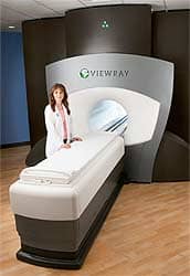
| “For the first time, we actually can see the area we are going to treat on CT scanners built into the treatment machines.” —Gordon L. Grado, MD Southwest Oncology Center |
The ideal radiation therapy protocol delivers a killing dose to the tumor and a zero dose to normal tissues. Many biological phenomena stand in the way of achieving this ideal. Most obviously, the patient may not be in the same position when the treatment is delivered as when the plan was created (interfraction error). Customized immobilization devices can reduce this error, as does ensuring correct alignment of bony landmarks at the start of a session.
However, these measures do not guarantee on-target radiation delivery, because cancers move (intrafraction error). This statement is obvious when the cancer is a lung tumor, which is displaced by breathing and by the motion of the heart. But it also is true for a less-obviously mobile lesion, such as a prostate cancer, which will be shifted by a filling bladder and progression of materials through the intestines, propelled by peristalsis. Moreover, with time and treatment, the radiation target may change both its size and its shape. For example, in women undergoing radiation therapy for uterine cervical cancer, the target volumes shrank an average of 27% by the third week of treatment,1 and in patients with gastrointestinal cancers, at least one fourth of the lesions moved 5 mm or more in one direction or another between the planning session and treatment delivery.2 A few cancers, such as those in the prostate, can be implanted with metal (for example, gold) seeds for pretreatment (or even intrasession) relocalization,3 but most tumors are not accessible for this maneuver.
How can the radiation therapist compensate for all this movement? If a lung tumor can move as much as 4 cm (a possible superoinferior range of motion, according to clinical studies), the only alternative has been to include a large margin of normal tissue in the radiation beam to ensure coverage of the entire tumor. As a result of this demand for large fields, the morbidity of radiation therapy for some tumors was legendary, and the tumor dose sometimes had to be reduced, increasing the risk of local recurrence. Conformal radiation and intensity-modulated radiation therapy (IMRT) were early steps in focusing the radiation beam precisely, and now there has been another step toward the goal: image-guided radiation therapy (IGRT). Here, the target is relocalized each day immediately before radiation delivery on the basis of internal soft-tissue anatomy rather than bony structures or skin tattoos. One result has been an escalation of the requirement of imaging capability in the radiation therapy suite.
Attempts have been made toward image guidance in the past. A popular technique was a megavoltage radiograph. Sometimes, this was printed on film, a time-consuming step. Easier to use was the electronic portal imaging device (EPID). Another option was planar kilovoltage imaging. Unfortunately, none of these methods in their original iterations yielded good soft-tissue—and, thus, target—images. A step forward was linear accelerators incorporating cone-beam CT technology with megavoltage or kilovoltage beams and a flat-panel detector. Today, radiation therapy departments are acquiring powerful CT scanners with excellent soft-tissue resolution for planning and delivery that are linked to the linear accelerator. Such purchases mark a dramatic change in the way radiation therapy departments are equipped.
New Demands on CT Scanners
“In the past,” recalls Gordon L. Grado, MD, of the Southwest Oncology Center (Scottsdale, Ariz), “radiation oncologists were often stuck with equipment that the diagnostic radiologists no longer wanted when they upgraded to the best machine available. So the therapists had, perhaps, a single-slice hand-me-down CT scanner that took half an hour to get all the images necessary for therapy planning or patient setup.”
For a growing number of departments, this is no longer acceptable. IGRT demands cutting-edge CT scanners for planning and execution.
“For the first time, we actually can see the area we are going to treat on CT scanners built into the treatment machines,” Grado says in describing the IGRT system he uses. “We can compare the CT images we obtained for planning with the scan that we obtained the day of treatment and fuse those images with the dosimetry information. As a result, we know where the radiation fields should be in that patient on that day. We can optimize patient position, be sure the blocks used to protect vital structures actually are protecting them, and ensure that the tumor is treated to the planned dose.”

|
| Aquilion Large Bore (LB) from Toshiba America Medical Systems boasts the industry?s largest bore opening of 90 cm and a 70-cm acquired field-of-view. |
The multislice CT scanners being used for IGRT have some special features. Most prominently, they have wide bores that allow the patient to be examined in the treatment position. For example, a woman being treated for breast cancer can be arranged on a board with a 25° tilt and with her arms above her head, or a patient being treated for a pelvic tumor can be placed in the dorsal lithotomy position. The LightSpeed RT16 and LightSpeed Xtra from GE Healthcare (Waukesha, Wis) have an 80-cm gantry with a 65-cm field of view (FOV). Also available from GE Healthcare is the Discovery ST scanner, capable of positron emission tomography (PET) as well as CT. The demand for PET has grown, as it has been found that the metabolic study leads to revision of almost half of radiation treatment plans.4 The GE Healthcare scanners can be equipped with a treatment planning couch insert that allows routine replication of exact patient positions. The Aquilion LB from Toshiba America Medical Systems (Tustin, Calif), has a 90 cm gantry and uses the new QuantumPLUS detector that has an additional 98 channels in the x, y direction, providing a true scan FOV of 70 cm. This 16-slice scanner permits true cone-beam imaging, using a proprietary algorithm based on the modified Feldkamp reconstruction. Optional fluoroscopy can display three consecutive images simultaneously. The Primatom integrated CT scanner and linear accelerator from Siemens Medical Solutions (Malvern, Pa), with an 82-cm gantry and a 50-cm FOV, incorporates the Beamview electronic portal imaging device. The company also offers the Biograph PET/CT scanner for oncology.

|
| Figure 1. Images of 3 mm polyp in colon provide a clinical example of improved geometric accuracy with thinner slices. Image courtesy of Campus Charite Mitte, Berlin, Germany. |
All of these scanners are capable of capturing submillimeter slices (for example, 0.5 mm for the Aquilion LB) with excellent soft-tissue contrast, making contrast studies unnecessary in most cases and, thus, improving patient safety and comfort while reducing procedure time and cost. The importance of thin slices for precise tumor localization and contouring can be seen in Figure 1.
These wide-bore scanners also are suitable for most obese patients, a population becoming increasingly common. “With IGRT, you need a space between the patient’s skin and the FOV, because if you cut off a piece of the tissue from the image, the dosimetry is not going to be correct,” Grado points out. “At Southwest Oncology, we have had some of the biggest patients I have ever seen in my life, and we have not had problems with targeting.”
To exploit the large number of images acquired, the scanners are supported by high-capacity workstations and specialized software capable of 3D reconstructions, contouring throughout the volume of the tumor, and image fusion. Of special value is the 4D (time-resolved) capability, essential for the targeting of lung cancers. Images are acquired throughout the respiratory cycle, or retrospectively, in which images are acquired throughout the cycle and sorted (binned) into the various phases in the cycle with the aid of respiratory timing information. If the patient does not breathe regularly on request, audio or video coaching may be provided. In patients with compromised pulmonary function, modified ventilator equipment may be employed for active breath control.4 The same method is then used during radiation delivery.
Wide-Bore Interest Widens |
|
Wide-bore scanners used in conjunction with image-guided radiation therapy systems are attracting interest from diagnostic radiologists serving other departments of the hospital. For example, it may be difficult or risky to position a trauma patient to enter a standard-size scanner, so this patient may be examined on the radiation therapy department’s scanner, located in the radiology department. A whole-body scan with 2-mm slices can be acquired in less than 20 seconds. The wide bore also appeals to interventional radiologists needing access to the patient’s anatomy as well as real-time images. But perhaps the greatest demand is to accommodate America’s widely publicized growing girth. Michael MacLeod, product manager of CT oncology at Toshiba America Medical Systems (Tustin, Calif) and a former hospital radiology department administrator, recalls some of the problems of scanning obese patients. “Many times, we could just barely fit them into a 70- or 72-cm scanner,” he recalls. “Even when we did, our problems were not over, because the field of view on those instruments was only 50 cm, so a lot of information was missing. This also contributed to out-of-field artifacts, which made the in-field image quality worse.” |
As an example of the capabilities of the image-analysis software, Paul Anderson, general manager oncology for GE Healthcare, describes their Advantage Sim MD package. “In a single session of 10 or 15 minutes, it is possible to create a treatment plan based on a standard dosimetry scan, with a four-dimensional thin-slice CT data set containing as many as 2,000 images, that can be registered simultaneously to PET and MR images. The radiation therapist can contour a moving tumor and watch the tracing being updated also on PET and MR scans to make certain that all of the involved areas are included and that nontargets, such as the kidneys, are excluded.” Also available is Advanced Lung Analysis, which permits automated segmentation of lung nodules, comparison of multiple examinations of the same patient, and displays of segmented nodule outlines on axial images.
Does launching IGRT require you to erect a new building to accommodate the equipment? Maybe not. About half of today’s cancer centers, especially those doing IMRT, already have some type of CT simulator, and for them, there would be no space change, only a technology change. Even late adopters have real estate for conventional x-ray simulators that can be converted to accommodate a new CT scanner.
Evidence of Success
The clinical history of IGRT is too short to allow determination of whether it cures more cancers, but it clearly reduces treatment morbidity.
“With image-guided radiation therapy, what we don’t want to treat, we don’t treat,” Grado notes. “Now when I plan, I don’t think, ?I want to treat the pelvis.’ Instead, I think, ?I want to treat a particular lymph node group.’ The small bowel and other normal tissues can be shielded as never before.
“Insurance companies do not recognize these benefits of IGRT to the degree they should,” he continues. “As an example of the reduction in morbidity that can be achieved, we treated the entire para-aortic lymph-node chain of a man with extensive prostate cancer, and his only complaint was low energy. In the past, there probably would have been severe colorectal problems, and he would have spent a lot of money on expensive palliative medications. With IGRT, if he had lived near our center, he would have been able to work the whole time he was receiving radiation therapy.”
The Future

|
| GE Healthcare?s LightSpeed RT16 has an 80-cm gantry with a 65-cm field-of-view. |
Radiation therapists already are moving on to biological image-guided radiation therapy, or what Ajit Singh, PhD, president of oncology care systems at Siemens Medical, has described as “the marriage of molecular imaging and oncology.” The goal is to define precisely the outer edge of the tumor and to overcome the problem of identifying any residual tumor in the presence of postradiotherapy scar. One molecular method is PET, which is expected to be refined by the introduction of cancer-specific tracers now in development, such as radiolabeled antisense molecules and antibodies.4 A particularly promising option is 18F-fluorothymidine, which is taken up preferentially by tumors because of their greater synthesis of DNA.4
The future may see even more use of imaging. Early work is being done in adaptive radiation therapy, in which the beams may be retargeted during a treatment session, if appropriate. Even if this maneuver does not become common, radiation therapy departments likely will need high-capability CT scanners. Radiation oncologists will demand it—and so will their patients.
Judith Gunn Bronson, MS, is a contributing writer for Medical Imaging.
References
- Lee JE, Han Y, Huh SJ, et al. Interfractional variation of uterine position during radical RT: weekly CT evaluation. Gynecol Oncol. August 17, 2006. Available at: www.sciencedirect.com Accessed September 11, 2006.
- Perkins CL, Fox T, Elder E, Kooby DA, Staley CA III, Landry J. Image-guided radiation therapy (IGRT) in gastrointestinal tumors. JOP. 2006;7:372?381. Available at: www.joplink.net/prev/200607/06_c.html. Accessed September 11, 2006.
- Sorcini B, Tilikidis A. Clinical application of image-guided radiotherapy, IGRT (on the Varian OBI platform). Cancer Radiother. July 31, 2006. Available at: www.ncbi.nlm.nih.gov Accessed September 11, 2006.
- Xing L, Thorndyke B, Schreibmann E, et al. Overview of image-guided radiation therapy. Med Dosim. 2006;31:91?112. Available at: www.ncbi.nlm.nih.gov Accessed September 11, 2006.
Additional Reading
- Heron DE, Smith RP, Andrade RS. Advances in image-guided radiation therapy: the role of PET-CT. Med Dosim. 2006;31:3?11.
- Jaffray DA. Emergent technologies for 3-dimensional image-guided radiation delivery. Semin Radiat Oncol. 2005;15:208?216.
- Ma CM, Paskalev K. In-room CT techniques for image-guided radiation therapy. Med Dosim. 2006;31:30?39.
- Song WY, Schaly B, Bauman G, Battista JJ, Van Dyk J. Evaluation of image-guided radiation therapy (IGRT) technologies and their impact on the outcomes of hypofractionated prostate cancer treatments: a radiobiologic analysis. Int J Radiat Oncol Biol Phys. 2006;64:289?300.


