By Elaine Sanchez Wilson
With the push for integrative health and collaborative care, now more than ever radiologists are serving in roles that transcend pure diagnostician. In an oncology specialty that espouses a multimodality, team-based approach, imaging is poised to fulfill, and go beyond, clinical needs in the screening, diagnosis, staging, and surveillance of cancer patients.
“Radiologists are becoming major actors in cancer patients’ management, at every step of the disease,” said Eric de Kerviler, MD, a radiologist at Saint-Louis Hospital in Paris. “Before diagnosis, they participate in screening campaigns. At diagnosis, they now perform most of the biopsies, the first step in a patient’s history. Then, they are required to assess the therapeutic response in patients with disseminated disease. In patients with solitary or oligometastatic tumors, ablation techniques represent an effective local cancer control option. Lastly, they play an important role in end-of-life care strategies, with palliative procedures such as nerve block, vertebroplasty, or derivations.”
Edward Y. Kim, MD, assistant professor of radiation oncology at the University of Washington (UW) School of Medicine, pointed out that radiologists frequently work alongside clinicians in choosing the best imaging modality for a particular case. “If you look at the American Society of Clinical Oncology’s ‘Choosing Wisely’ campaign, three out of five of its recommendations center around the use of imaging,” he said. “Radiologists are valuable—and critical—members of a patient’s care team, and they have an opportunity to lead the way in selecting the most appropriate imaging.”
At this year’s meeting of the Radiological Society of North America, Kim will be discussing scientific presentations at the BOOST session on gastrointestinal malignancies. “Radiation Oncology and Radibiology (Gastrointestinal)” will take place on Wednesday, December 4, from 10:30 am until 12 pm in Room S104A.
Kim also mentored UW resident physician Laura Koller, MD, who will present findings from their study on “Solitary Fibrous Tumors: Radiographic and Pathologic Responses to Neoadjuvant Radiotherapy.” The researchers found that all of the study’s patients experienced a decrease in tumor volume after preoperative radiotherapy. While there was no direct correlation between the magnitude of response and the degree of necrosis on pathology, 80% of patients demonstrated treatment response. The poster presentation will take place on Thursday, December 5, from 12:45 to 1:15 pm in Room LL-ROS-TH3B at the Lakeside Learning Center.
In the next few pages, you will discover additional oncological imaging talks and topics to attend at RSNA 2013.
Molecular Imaging
Ronald Korn, MD, PhD, CEO and medical director of a research and imaging core laboratory, Imaging Endpoints, attends numerous conferences in a given year and praises the RSNA meeting as one of the most exciting medical conventions in the world. “I think it’s the world’s greatest marketplace for radiology,” he said. “It’s the marketplace for ideas, new trends, equipment, and networking. Often, the greatest conversations are those you have with colleagues in the hallways, finding out what they are up to and explaining what you are up to. What is really neat is the great arena of connections that the RSNA supplies.”
Korn agrees that radiologists are central to the care team of oncologists; in fact, Korn himself is a radiologist who serves as the medical director of the oncology program at the Virginia G. Piper Cancer Center in Scottsdale, Ariz. “The way I got here was basically becoming an oncologist myself through radiology,” Korn explained. “I have found that the way you can make yourself most valuable to the oncologist is to understand their specialty. You have to have a very detailed knowledge of molecular biology, you have to understand molecular oncology and new trends, and you have to speak their language. You become a critical part of the team when you really know what information they are seeking and how to get it in the most effective and efficient way using radiology.”
For the last several years, Korn has moderated RSNA’s Molecular Imaging Symposium. This year, the topic is “Preparing for tomorrow: The application of novel and advanced imaging in clinical oncology.” “The concept is how do you bridge the gap between the needs of the oncologist and the tools and capabilities of the radiologist,” Korn said. “What is critical to a strong relationship with oncologists is an understanding of how oncologists make treatment decisions and then translating that understanding into specific imaging recommendations to help in the decision-making process.”
According to Korn, a new generation of targeted therapies is currently in clinical trials or just starting to emerge in clinical medicine. His session on molecular imaging will touch upon how, in imaging, the response to these therapies is not necessarily shrinkage in tumor size but perhaps an alteration in its appearance. The talks also will go into detail on the regulatory approval process for PET and MRI tracers, which can help in optimizing treatment or in selecting patients who will respond to those treatments. Furthermore, they will explore the biomarker space, or “imaging at the genomic level” as Korn puts it, as well as pharmacodynamic imaging, which is “understanding what’s happening at the extracellular, blood flow, and even the enzymatic level, seeing how the dynamic processes of tumors are changing,” he said.
Korn’s session will take place on Monday, December 2, in Room S406B from 8:30 to 10 am. Topics will include “Fluorescence and Optoacoustic Imaging Heads to the Clinics,” “CT Biomarkers and How to Use Them,” “The Use of Novel PET Tracers: What is in the pipeline for approval?”, and “Systems Diagnostics: The Future of Diagnostic Medicine?”. Korn hand-selected the speakers, who he says represent key opinion leaders in the field. “I chose them because they have what I think are some of the most interesting concepts that are likely to make it into the clinic within the next 2 to 5 years,” he said.
Image-guided Biopsy
Other recent advances in oncology include personalized medicine strategies, which, according to de Kerviler, will impact patient care in two major ways. “The first is to be able to demonstrate receptors or mutations suitable for targeted therapies,” he said. “This necessitates optimal tissue sampling and management when a biopsy is performed. Obtaining a large amount of tissue with frozen specimens in different tumor areas is mandatory for molecular studies.
“The second is to find a reliable tool to monitor the therapeutic response when using noncytotoxic drugs,” he said. “RECIST (Response Evaluation Criteria in Solid Tumors) and WHO criteria clearly show their limitations with these new therapies, and we need new biomarkers. Choi criteria, microvascular parameters, diffusion studies, or PET may be combined to morphological criteria to better assess the therapeutic response.”
As one of four French speakers who will be presenting within the “France Presents” session, de Kerviler will discuss the improvement of the diagnostic yield of image-guided biopsies when combining morphological and functional imaging. “Intratumor heterogeneity accounts for a significant number of insufficient or inappropriate biopsy material,” he said. “Macroscopically, tumors appear as heterogenous masses, with foci of necrosis, fibrosis, so that a small amount of tissue may not adequately represent the most aggressive component.
“Functional imaging can provide a more complete insight into living tumors, demonstrating areas of increased metabolism, cellularity, or angiogenesis,” de Kerviler said. “Targeting biopsies toward the anomalies on functional images increases the chances to obtain representative cores of tissue.”
“Beyond Morphology: Molecular imaging for biopsy guidance in oncology” will take place on Monday, December 2, from 10:55 to 11:15 am in Room E353C.
Dynamic Contrast-enhanced Ultrasound
Agreeing that molecular imaging for personalized medicine is an emerging trend in oncology, Nathalie Lassau, MD, PhD, noted that her imaging laboratory at the University of Paris will be launching a program in this area of research.
Meanwhile, at this year’s meeting, Lassau will present the final results of a multicentric study on dynamic contrast-enhanced ultrasonography (DCE-US) that was supported by the French National Institute of Cancer. The study, which included 19 French hospitals, 65 radiologists, and 539 patients, aimed to demonstrate the feasibility of DCE-US and determine the best parameters and timing for evaluating response to therapies. Researchers confirmed that the DCE-US is the first functional imaging technique to give validated parameters predictive of tumor progression in a large multicenter cohort. Moreover, the indication was implemented in the world guidelines as good clinical practice recommendations.
Lassau recognized that DCE-US, which is widely used in Europe, stands to enhance radiologists’ value to the cancer patient’s care team. “This technique reinforces the link between radiologists and patients because we discuss in real time the results before consultation with the oncologist,” she said, adding that the radiologist-patient relationship is strengthened through the opportunity for discussion.
“Validation of Best Surrogate Markers of DCE-US to Predict PFS for Different Anti-angiogenic Treatments” will be presented on Monday, December 2, from 10:40 to 10:50 am in Room E451A.
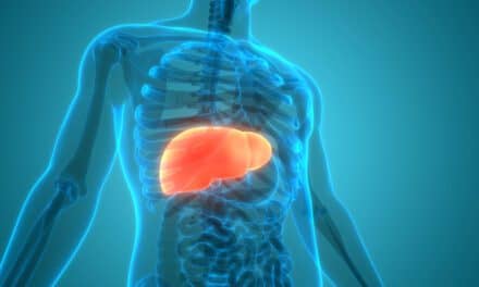
Hepatocellular Carcinoma Detection and Treatment
Michele Di Martino, a PhD candidate at Sapienza University of Rome, is set to have a busy RSNA meeting. She will be speaking in three presentations: two on hepatocellular carcinoma (HCC) and one on the quantification of liver steatosis in children. Specifically, one of the lectures on hepatocellular carcinoma compares CT and MRI with a liver-specific contrast agent and the explanted liver. The second talk compares RECIST 1.1 criteria and mRECIST criteria in the follow-up of patients with advanced HCC treated with antiangiogenetic drugs.
According to Di Martino, the detection of HCC in cirrhotic liver is vital in determining the stage of disease and the correct treatment, and if it is detected early enough, therapeutic strategies such as liver resection, liver transplant, radiofrequency ablation, and chemoembolization may be available. While the diagnosis of HCC typically requires the histologic confirmation, specific imaging patterns may allow a noninvasive diagnosis based solely on CT and MRI examination, thereby avoiding an invasive biopsy.
“Several published papers have already investigated the role of CT and MRI in the detection of hepatocellular carcinoma in cirrhotic patients but only a few works have compared them directly with the explanted liver, which is the best gold standard,” she said. The researchers ultimately found that MRI with liver-specific contrast agent, performed with state-of-the-art equipment, seemed to be the best imaging modality for the detection of HCC in cirrhotic patients.
Di Martino also will present findings from a study comparing RECIST 1.1 criteria with mRECIST criteria in the evaluation of radiological response to therapy in patients treated with the antiangiogenetic drug Sorafenib. Their aim was to evaluate which patients could benefit from Sorafenib, which had been introduced in the American Association for the Study of Liver Diseases (AASLD) guidelines for the treatment of cirrhotic patients with advanced hepatocellular carcinoma. Two clinical trials, SHARP and Asia-Pacific, have already demonstrated that patients treated with Sorafenib significantly extended their life (by an average of 3 months) compared with patients treated with a placebo.
“Despite this relevant clinical finding, in both trials a poor radiologic response to treatment has been noticed,” she said. “In our experience, mRECIST criteria help to better stratify patients who are usually considered as having? stable disease with RECIST 1.1 criteria. In particular, patients who respond to Sorafenib should benefit from this treatment. By contrast, patients with stable or progressive disease should stop therapy.”
“Detection of Hepatocellular Carcinoma (HCC) in Liver Transplant Candidates: Intraindividual Comparison of Gadobenate Dimeglumine (Gd-BOPTA) Enhanced MR Imaging and Multiphasic 64-slice CT” will be held on Monday, December 2, from 11:30 to 11:40 am in Room E353A. “Accuracy of mRECIST versus RECIST 1.1 in Predicting Outcome in Hepatocellular Carcinoma Treated with Sorafenib” will take place Monday, December 2, in Room E451A during the session that runs from 10:30 am until 12 pm. “Quantification of Liver Fat Content in Adolescents with Non-alcoholic Fatty Liver Disease: Comparison of Triple-Echo Chemical Shift Gradient-Echo Imaging and In Vivo Proton MR Spectroscopy” will take place on Tuesday, December 3, in the session that runs from 3 to 6 pm in Room S102AB.
###
Elaine Sanchez Wilson is a contributing writer for Axis Imaging News.


